Charcot-Marie-Tooth (CMT) disease is a hereditary, progressive peripheral nerve disorder that introduces intricate neurological and musculoskeletal challenges to a patient’s lower extremities. Characterized by a slow degeneration of the motor and sensory nerves traveling to the feet and shins, CMT leads to classic muscle imbalances. This progressive muscle wasting typically causes a distinct combination of a rigid high arch (pes cavus), tightly clawed toes, and severe flaccid foot drop. For individuals navigating this complex condition, implementing specialized cmt disease orthotics is a vital step toward preserving daily mobility. This comprehensive case review details a multi-disciplinary treatment approach for a CMT patient experiencing chronic ankle instability and frequent falls, showcasing how a custom-engineered triplanar ankle-foot orthosis successfully restored alignment, rebalanced walking forces, and returned the patient to a safe, independent lifestyle.
Clinical Case Presentation and Gait Baseline
The patient, a 34-year-old female accountant, presented for a comprehensive orthotic evaluation with a formal diagnosis of CMT Type 1A. Over the past two years, she reported a significant decrease in her walking stability, a loss of balance in low-light environments, and an average of three dangerous falls per month. A physical assessment revealed severe muscle thinning across the calf muscles, giving her legs a classic “inverted champagne bottle” appearance. Her feet exhibited a rigid pes cavus structure with severe clawing across all toes. Manual muscle testing scored her ankle dorsiflexors at a low two out of five, resulting in a flaccid foot drop. When attempting to walk without support, she demonstrated an exhausting steppage gait, lifting her knees abnormally high while her heel tilted sharply inward, placing her at risk for repetitive lateral ankle sprains.
Engineering the Custom Triplanar AFO
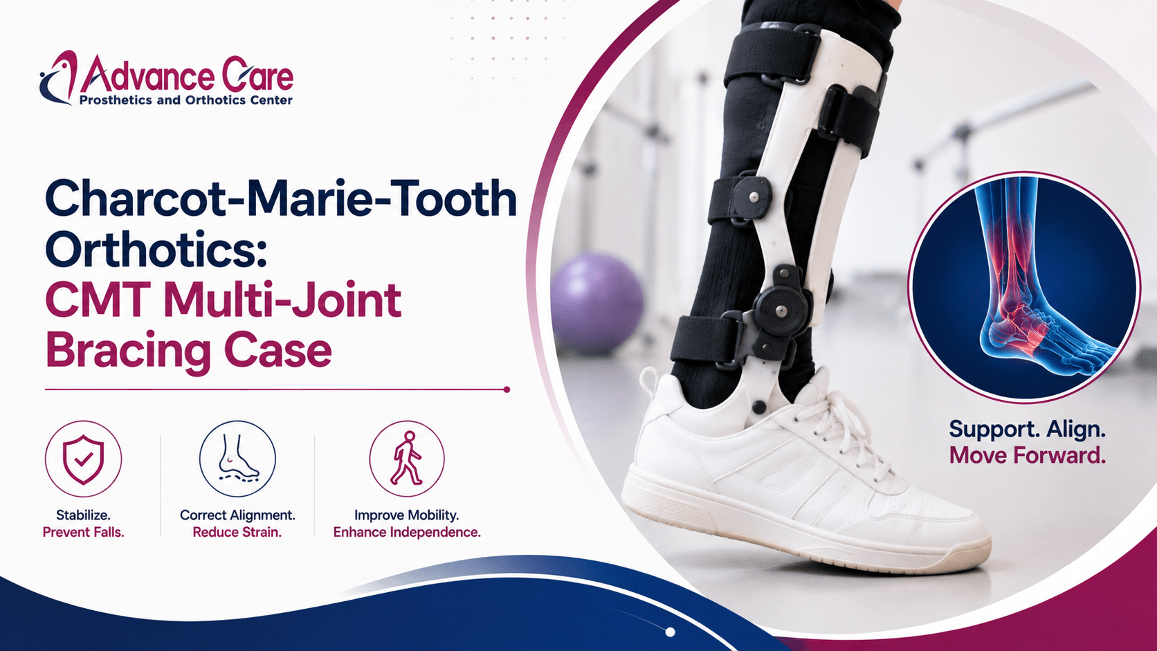
To safely manage her multi-joint instability and control forces across all three planes of movement, the clinical team designed a high-performance, custom-molded carbon composite ankle-foot orthosis.
- Sagittal Plane Control (Foot Drop): The brace incorporates a rigid posterior strut that holds the ankle securely at a neutral ninety-degree angle during the swing phase, instantly eliminating toe drag and the need for a high steppage gait.
- Coronal Plane Alignment (Heel Inversion): The custom carbon shell features an extended inner wall and a deep heel cup that grasps the calcaneus bone, counteracting the inward varus tilt and stabilizing the lateral ankle ligaments.
- Transverse Plane Support (Forefoot Adduction): The custom-molded footplate is crafted with full total-contact support that arches up to fill the high pes cavus gap, distributing body weight evenly across the entire sole.
Multidisciplinary Rehabilitation Protocol Matrix
Restoring the patient’s walking confidence required a highly coordinated, twelve-week program combining advanced custom bracing with targeted daily physical rehabilitation.
| Recovery Component | Clinical Focus and Interventions | Biomechanical and Neurological Goal |
| Phase 1: Adaptation | Gradual break-in schedule; wearing the brace for 1-2 hours at home; skin check training. | Prepares skin tissues for structured contact; builds early confidence in the brace support. |
| Phase 2: Core Strengthening | Targeted exercise targeting the proximal hip flexors, gluteal muscles, and core stabilizers. | Rebalances muscle groups above the brace; improves overall posture and walking balance. |
| Phase 3: Balance Training | Proprioceptive training using balance boards; navigating ramps, stairs, and uneven grass. | Stimulates remaining nerve pathways; eliminates fear of falling during community activities. |
Quantifiable Objective Outcomes
Following twelve weeks of consistent, integrated care, the patient returned for a follow-up assessment using standardized clinical outcome metrics to evaluate her progress. Her walking velocity on the 10-Meter Walk Test improved from a baseline of 0.51 meters per second up to an efficient 0.96 meters per second, allowing her to cross streets safely during community commuting. Her scores on the Berg Balance Scale rose from 38 out of 56 up to a secure 51 out of 56, moving her out of the high fall risk category. Most importantly, the patient reported a complete elimination of falls during her daily work and personal routines, along with a significant reduction in lower back and hip fatigue.
Frequently Asked Questions
Why does CMT disease cause the lower leg muscles to thin out into an inverted bottle shape?
CMT damages the peripheral nerves that supply the lower leg, disrupting the vital electrical signals needed to keep muscles working. Over years of reduced nerve input, the calf and shin muscles thin out permanently, while the upper thigh muscles typically retain their normal strength.
Can custom CMT orthotics stop the underlying genetic disease from progressing?
No, orthotic braces cannot alter or halt the underlying genetic nerve progression of CMT. Instead, they act as essential biomechanical tools that manage the physical consequences of the disease, preventing bone deformities, stabilizing weak joints, and keeping you safely mobile.
Is it necessary to use a custom-molded brace rather than an off-the-shelf foot drop splint for CMT?
Yes, custom bracing is vital for CMT management. Generic off-the-shelf splints are designed for flat, flexible feet and cannot accommodate the rigid high arches or severe inward heel tilting characteristic of a CMT foot structure without causing painful pressure sores.
How often should a patient with CMT inspect their skin when breaking in a new brace?
During the initial two-week break-in period, the patient should remove the brace every two hours to inspect the skin carefully. Look for deep red marks over the heel or arch; if a red spot takes longer than twenty minutes to fade, the brace requires a quick adjustment by your orthotist.
What styles of shoes work best when pairing them with a custom carbon fiber AFO for CMT?
Look for supportive athletic sneakers or casual shoes with extra depth, a wide toe box to prevent claw toe irritation, a stable rubber outsole for traction, and a completely removable factory insole to create ample room for the brace.
Conclusion
The exceptional physical progress detailed in this case review underscores the vital importance of advanced biomechanical engineering and coordinated care in managing chronic neurological disorders. As demonstrated throughout real-world clinical reports, utilizing precision-tailored cmt disease orthotics offers an extraordinarily effective pathway for counteracting progressive muscle wasting, stabilizing an inward-rolling heel, and completely eliminating dangerous toe drag. By combining an advanced carbon fiber brace with a structured physical therapy program, you can successfully bypass muscle deficits and safeguard your long-term joint health. Consult with a dedicated team of neurologists, physical therapists, and certified orthotists to engineer your personalized orthotic care plan, helping you step forward with absolute safety, comfort, and independence.